Intra-vascular Imaging (OCT and IVUS)
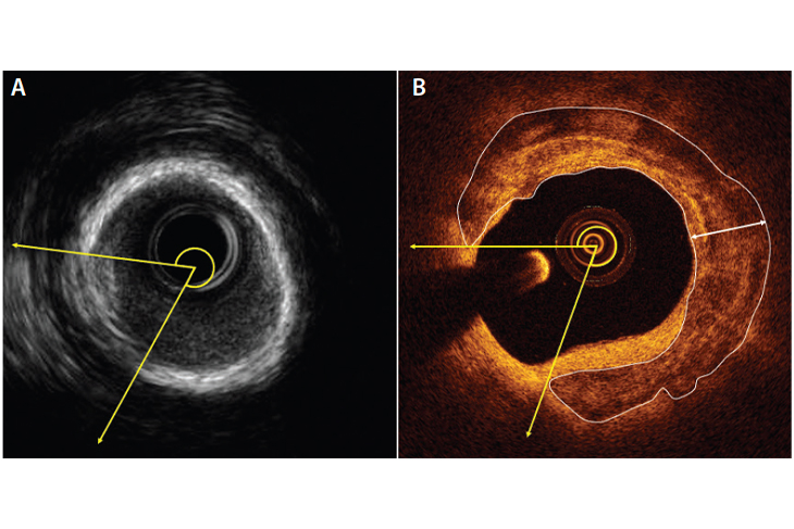
Intra-vascular imaging, such as Optical Coherence Tomography (OCT) and Intravascular Ultrasound (IVUS), are diagnostic procedures used to obtain detailed images of the interior of blood vessels and to assess the presence and severity of conditions such as coronary artery disease, peripheral artery disease, and aneurysms.
The procedures involve introducing a catheter into the blood vessel and using either ultrasound or light waves to obtain images of the blood vessel walls and surrounding tissue. Intra-vascular imaging can help guide treatment decisions, such as whether a patient should undergo percutaneous coronary intervention (PCI), coronary artery bypass surgery, or other treatments.
Eligibility for intra-vascular imaging depends on several factors including the presence and location of conditions affecting the blood vessels, the patient's overall health, and the presence of other medical conditions.