Contrast Echo

What is Contrast Echo?
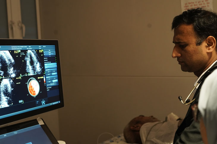
Contrast echocardiography is a non-invasive technique used to take pictures of the heart while it's functioning. It gives pictures of the heart with more quality and accuracy. It will help the cardiologist to examine the working condition of the cardiac muscles and valves and the size of the heart chambers.
How is it performed?
Before the contrast echo, a dye is injected into the veins of the patients. This dye will make the heart muscles and other structures more visible and understandable during the ultrasound. The machine uses ultrasound waves to picture the heart with dye in it. The procedure of contrast echo usually takes around 30 minutes. The doctor will place an IV line into your vein on the arm. A gel is applied on the chest, and the transducer, which emits the sound waves, is placed on the chest. The sound waves produced by the transducer transmit back from the heart, and these sound waves will be recorded by the machine.


